In your eyes: Clues to heart disease risk?
Changes both around and inside your eyes — some visible only with special tests — may be harbingers of heart disease.

Are the eyes a window to the heart as well as the soul? New insights and older observations suggest the answer is yes.
Sudden vision changes such as blurriness, dark areas, or shadows could be a blockage in an eye blood vessel, which can foreshadow a more serious stroke in the brain. And growing evidence hints that subtle, early damage to tiny blood vessels in the eyes may predict cardiovascular disease. Other unusual eye changes also can be clues to possible heart problems, such as small, yellowish bumps around the eyes, or pupils that widen and constrict with the heartbeat (see "A pulsating pupil and a leaky heart valve?").
A pulsating pupil and a leaky heart valve?When the heart’s aortic valve doesn’t close tightly (a condition called aortic regurgitation), small amounts of blood leak backward into the heart instead of being pumped into the body. Symptoms typically include fatigue and breathlessness. In severe cases, another possible sign is a rarely documented eye phenomenon: pupils that dilate and constrict in synchrony with the heartbeat. Called Landolfi’s sign, it was first described by Italian physician Michele Landolfi in 1909. Compared with people who have normal hearts, people with aortic regurgitation have a wide pulse pressure, which means there is an unusually big difference between the two numbers in their blood pressure readings (that is, the systolic and diastolic numbers). The pupil pulsations reflect these large pressure fluctuations. |
Retina changes reflect heart health
A network of blood vessels, each no wider than a strand of hair, supply blood to the retina, the light-sensitive tissue at the back of the eyeball. "Like arteries throughout the body, these tiny vessels can be damaged by chronic conditions such as diabetes and high blood pressure," says Dr. Nimesh Patel, an ophthalmologist who specializes in retina disorders at Harvard-affiliated Massachusetts Eye and Ear hospital.
Over time, high blood sugar (the hallmark of diabetes) causes the walls of vessels supplying the retina to weaken and leak fluid into the surrounding tissue. This condition, called diabetic retinopathy, can progress, causing further damage that may impair vision. Poorly controlled high blood pressure can cause retinal arteries to narrow or break and bleed into the retina.
While both diabetes and high blood pressure are well-known contributors to heart disease, people may not know they have these conditions if they haven’t seen a primary care doctor in years, says Dr. Patel.
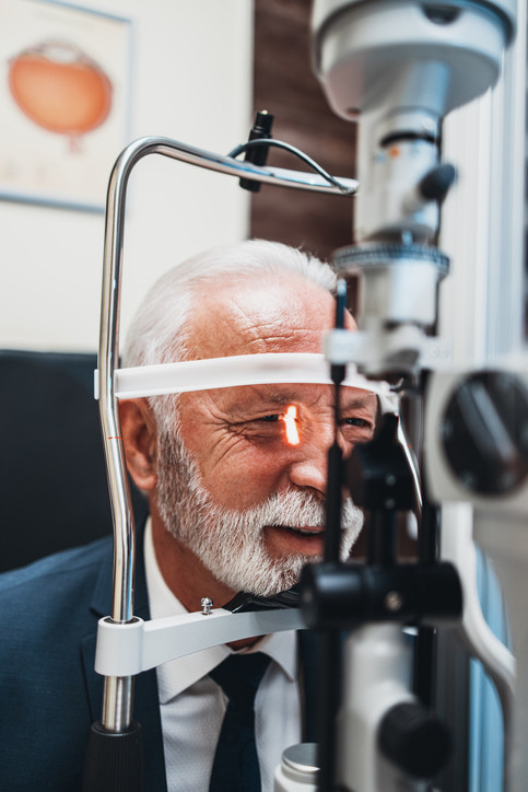
"Sometimes, patients come in with vision problems and a retinal photo or exam shows a piece of plaque blocking a retinal artery," he says. Known as a retinal artery occlusion, this condition indicates a greater risk of a stroke in the brain. Blood clots may also form in the veins that carry blood away from the retina. Both conditions — a retinal artery or vein occlusion — are referred to as eye (ocular) strokes.
People who experience eye strokes need further testing, including ultrasounds of the neck to check for a buildup of fatty plaque inside the carotid arteries and of the heart to check for blood clots.
More subtle changes seen in retina photos may foretell future heart problems. By analyzing the photos with machine learning (combined with health data such as age and blood pressure), several teams of researchers have created algorithms to predict a person’s risk of heart attack or stroke. At this point, the models still need to be improved and validated. But advances in both imaging and artificial intelligence mean such tests could be available in the not-too-distant future.
Yellow bumps around the eyes: Xanthelasmas
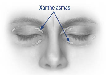
Some middle-aged and older adults develop soft, yellowish, cholesterol-filled bumps on or around their eyelids, most commonly near the nose (see illustration). Called xanthelasmas, these small growths aren’t painful and rarely affect vision. But they can be a sign of high levels of cholesterol, triglycerides, or other fats (lipids) in the blood. They’re somewhat more prevalent in women than men. People who have them should be sure to get a cholesterol test, also known as lipid test or lipid profile.

Illustration by Scott Leighton
Xanthelasmas are more common in people with genetic disorders that cause abnormally high cholesterol levels, such as familial hypercholesterolemia, which can cause LDL cholesterol levels of 190 or higher. (A healthier LDL is under 100.)
If your LDL is elevated, regular exercise and a high-fiber, plant-based diet can help lower it. Many people need cholesterol-lowering drugs such as statins, which will sometimes but not always reduce the size of a xanthelasma.
Half of people with xanthelasmas have normal lipid levels, and the growths are simply a cosmetic problem. But people with a family history of early heart disease should make sure their physician know about this possible sign of heightened cardiovascular risk.
Eye care advice
The American Academy of Ophthalmology recommends:
- People ages 40 to 64 who are otherwise healthy should get a complete eye exam from an ophthalmologist once every two to four years.
- After age 65, you should receive a complete eye exam every one to two years.
- People with type 2 diabetes should get a complete eye exam at the time of diagnosis and then annually after that.
About the Author

Julie Corliss, Executive Editor, Harvard Heart Letter
Disclaimer:
As a service to our readers, Harvard Health Publishing provides access to our library of archived content. Please note the date of last review or update on all articles.
No content on this site, regardless of date, should ever be used as a substitute for direct medical advice from your doctor or other qualified clinician.


