X-ray may be best screening tool for diagnosing knee pain
In the Journals

Image: © endopack /Thinkstock
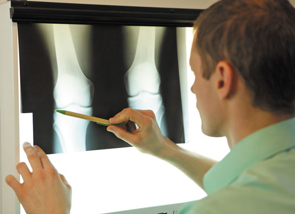
One of the most common reasons for a doctor's office visit is knee pain or injuries from osteoarthritis. While magnetic resonance imaging (MRI) is widely used by doctors to diagnose problems like torn knee ligaments and cartilage, a study in the September 2016 issue of the Journal of the American Academy of Orthopaedic Surgeons found that a simple x-ray may be a better diagnostic tool as it helps reduce time and cost.
The study looked at 100 MRI scans of knees from patients ages 40 and older and found that the most common diagnoses were osteoarthritis (39%) and meniscal tears (29%), which involve the tearing of the wedge-shaped pieces of cartilage in the knee joint. Almost 25% of MRI scans were taken before the patient first obtained an x-ray, and yet only one-half of those scans contributed to a patient's diagnosis and treatment for osteoarthritis.
Whether a patient needs surgery for knee problems depends on the amount of arthritis. If an x-ray shows that a person has significant arthritis, the MRI findings, like a meniscus tear, are less important because the amount of arthritis drives the treatment, according to lead researcher Dr. Muyibat Adelani of Washington University in St. Louis.
"X-rays are an appropriate screening test for knee pain in older patients, and often the results of an x-ray can tell whether an MRI would be even helpful," she says. In addition, an MRI costs about 12 times that of an x-ray (based on Medicare rates) and can take an hour to perform.
Disclaimer:
As a service to our readers, Harvard Health Publishing provides access to our library of archived content. Please note the date of last review or update on all articles.
No content on this site, regardless of date, should ever be used as a substitute for direct medical advice from your doctor or other qualified clinician.


