Diseases & Conditions Archive

Articles

Blood Testing

What Is It?

Blood tests enable doctors to assess your health by analyzing cells, chemicals, proteins and other substances in your blood. Some tests are recommended regularly to see if blood levels of certain cells or chemicals fall within a normal range. Others are done to help diagnose health conditions, such as allergies, anemia, and diabetes.

There are two typical methods for taking a blood sample. One, called venipuncture, involves drawing a vial of blood from a vein, usually on the inner surface of your arm near your elbow. The other, called a finger stick, is done by pricking your finger with a sharp blade to obtain a small amount of blood from a capillary. The method used depends on how much blood is needed for the test you are having.

Bone Marrow Transplant

What Is It?

A bone marrow transplant is a procedure used to treat certain types of cancer and some other diseases. Before the bone marrow transplant takes place, a person's bone marrow cells are destroyed with radiation or chemotherapy.

The cells that normally live in the bone marrow and that are responsible for making blood cells are then replaced. Bone marrow cells are blood cells that are located in the spongy center of bones. These include:

Bone Scan

What Is It?

A bone scan uses radiation to make images showing areas of bone where cells are unusually active. Unusually active cells can indicate cancer, bone trauma, infection or other disorders.

First, a radioactive chemical called an isotope is injected into a vein. The isotope enters the bloodstream and travels to the bones, where it emits gamma rays, which are similar to X-rays. A gamma camera can detect gamma rays. A computer analyzes the rays and forms an image (or scan) of the bones. Areas with potential problems send out more intense rays and appear as bright spots on the scan.

Carotid Ultrasound (Carotid Doppler)

What is the test?

Ultrasound uses sound waves instead of radiation to generate snapshots or moving pictures of structures inside the body. This imaging technique works in a manner similar to radar and sonar, developed in World War II to detect airplanes, missiles, and submarines that were otherwise invisible. After coating a small area of your skin with a lubricant to reduce friction, a radiologist or ultrasound technician places an ultrasound transducer, which looks like a microphone, on your skin and may rub it back and forth to get the right view. The transducer sends sound waves into your body and picks up the echoes of the sound waves as they bounce off internal organs and tissue. A computer transforms these echoes into an image that is displayed on a monitor.

Doppler ultrasound is a variation of this technique that not only shows internal structures but also examines the flow of blood through blood vessels. Using the Doppler effect—the change in the frequency of sound or light waves as they bounce off a moving object—this kind of ultrasound produces an image of blood in motion.

Cholecystectomy

What Is It?

Cholecystectomy is the surgical removal of the gallbladder, the small saclike organ located near the liver in the upper right side of the abdomen. It is attached to the main duct that carries bile from the liver into the intestine. Bile helps your body to break down and absorb fats. The gallbladder temporarily stores bile from the liver. When you eat, the gallbladder contracts, and squeezes extra bile into the intestine to aid digestion.

There are two ways to remove the gallbladder:

  • Traditional surgery - The surgeon cuts open the abdomen and removes the gallbladder through an incision that is about 6 inches long. The abdomen is then stitched closed again.
  • Laparoscopic surgery - The surgeon makes four small (less than an inch) incisions for a laparoscope and instruments. A laparoscope is a tube-like instrument with a camera for viewing, and with it the surgeon can to guide the surgical instruments to remove the gallbladder. The gallbladder is cut away from the liver and the bile duct and removed through one of the small incisions.

Over 90% of the time, laparoscopic surgery is used because it requires a shorter hospital stay, is less painful, and has a shorter recovery time compared to traditional surgery. Traditional surgery is usually used because the person has significant abdominal scarring from prior surgery, severe inflammation, unusual anatomy, or other factors that make surgery with a laparoscope very difficult and riskier.

Corneal Transplant

What Is It?

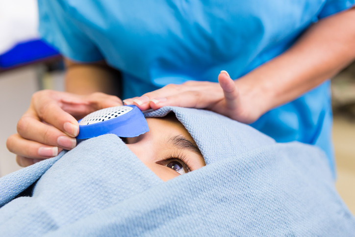
The cornea is the clear, round, "window" of tissue that allows light to enter the front of the eye. If the cornea becomes severely diseased or damaged, it can distort or even block the normal path of light into the eye. When this happens, light does not focus normally on the retina, the layer at the back of the eye that is responsible for sight. As a result, there can be a significant loss of vision in the affected eye.

When corneal conditions cause serious, irreversible vision problems, a corneal transplant often is the best solution. In a corneal transplant, an eye surgeon first removes the diseased or damaged area of cornea. The removed tissue then is replaced by a section of healthy cornea that has been taken from the eye of a dead donor.

Cystoscopy

What Is It?

Cystoscopy is a procedure that allows doctors to look inside the bladder and the urethra, the tube that carries urine from the bladder. A cystoscope is a tube-like instrument with lenses, a camera and a light on one end and an eyepiece on the other. With a cystoscope, your doctor can examine the urethra and the lining of the bladder. If necessary, your doctor can pass surgical instruments through the cystoscope to perform specific procedures. In most cases, a simple cystoscopy lasts 5 to 10 minutes. Procedures that are more complex take longer.

What It's Used For

Cystoscopy checks inside the bladder for tumors, sites of bleeding, signs of infection, stones (calculi) and causes of bladder outlet obstruction. It also can be used to:

Electroencephalogram (EEG)

An electroencephalogram (EEG) is a recording of the brain's electrical activity. Metal electrodes attached to the skin on the outside of the head transform electrical activity into patterns, commonly called brain waves. A polygraph machine records the brain waves. In some cases, the waves are transmitted to a computer screen. A basic EEG takes about 45 minutes, with a range of 30 minutes to 90 minutes.

Lightweight EEG devices allow people to walk around and perform normal daily activities while the devices detect and record brain waves over longer periods of time.

Endoscopy

What Is It?

Endoscopy describes many procedures that look inside the body using some type of endoscope, a flexible tube with a small TV camera and a light on one end and an eyepiece on the other. The endoscope allows doctors to examine the inside of certain tube-like structures in the body. Many endoscopes transmit the doctor's view to a video screen. Most endoscopes have attachments that permit doctors to take fluid or tissue samples for laboratory testing.

Upper endoscopy allows a doctor to see inside the esophagus, stomach and top parts of the small intestine. Bronchoscopy examines the large airways inside the lungs (bronchi). Sigmoidoscopy and colonoscopy examine different parts of the lower digestive tract. Each type of endoscopy uses a slightly different endoscope with a different name — an upper endoscope for upper endoscopy, a bronchoscope for bronchoscopy, a sigmoidoscope for sigmoidoscopy and a colonoscope for colonoscopy. Other endoscopes allow doctors to see inside the abdomen and inside joints through small incisions.

Heart Transplant

What Is It?

A heart transplant is surgery in which a patient with a life-threatening heart problem receives a new, healthy heart from a person who has died. In a heart transplant, the patient who receives the new heart (the recipient) is someone who has a 30 percent or greater risk of dying within 1 year without a new heart. Although there is no absolute age limit, most transplants are performed on patients younger than 70 years old.

The person who provides the healthy heart (the donor) is usually someone who has been declared brain dead and is still on life-support machinery. Heart donors are usually younger than 50, have no history of heart problems, and do not have any infectious diseases.

Free Healthbeat Signup

Get the latest in health news delivered to your inbox!

Sign Up
Harvard Health Publishing Logo

Thanks for visiting. Don't miss your FREE gift.

The Best Diets for Cognitive Fitness, is yours absolutely FREE when you sign up to receive Health Alerts from Harvard Medical School

Sign up to get tips for living a healthy lifestyle, with ways to fight inflammation and improve cognitive health, plus the latest advances in preventative medicine, diet and exercise, pain relief, blood pressure and cholesterol management, and more.

Harvard Health Publishing Logo

Health Alerts from Harvard Medical School

Get helpful tips and guidance for everything from fighting inflammation to finding the best diets for weight loss...from exercises to build a stronger core to advice on treating cataracts. PLUS, the latest news on medical advances and breakthroughs from Harvard Medical School experts.

BONUS! Sign up now and
get a FREE copy of the
Best Diets for Cognitive Fitness

Harvard Health Publishing Logo

Stay on top of latest health news from Harvard Medical School.

Plus, get a FREE copy of the Best Diets for Cognitive Fitness.

Harvard Health Publishing Logo

Stay on top of latest health news from Harvard Medical School.

Plus, get a FREE copy of the Best Diets for Cognitive Fitness.